Search for Human Egg-Cells; Identification, Features/Properties and Variance. https://www.ncbi.nlm.nih.gov/books/NBK26842/ (05-05-2021)
https://www.asme.org/wwwasmeorg/media/resourcefiles/shop/certification-accreditation/nmo-certification/nmo-certification_additional-information_notice-on-nuclear-scope-statements.pdf
https://www.springer.com/journal/41365/aims-and-scope
https://www.springer.com/journal/41365/aims-and-scope
https://inis.iaea.org/search/search.aspx?orig_q=RN:22017171
'The zona pellucida. (A) Scanning electron micrograph of a hamster egg, showing the zona pellucida. (B) A scanning electron micrograph of a similar egg in which the zona (to which many sperm are attached) has been peeled back to reveal the underlying plasma (more...)'
https://www.ncbi.nlm.nih.gov/books/NBK26842/
Scanning Electron Microscope, Micrography. Plasma detection. Yocto, nano, femto, pico, 100micro.
International system of units. STP.
Measures, Common Ratio (adjusted or not, calibrated, standardized, etc.), Equations, Formulaes.

Figure 20-21The zona pellucida
(A) Scanning electron micrograph of a hamster egg, showing the zona pellucida. (B) A scanning electron micrograph of a similar egg in which the zona (to which many sperm are attached) has been peeled back to reveal the underlying plasma membrane, which contains numerous microvilli. The zona is made entirely by the developing oocyte. (From D.M. Phillips, J. Ultrastruct. Res. 72:1–12, 1980.)

: Eggs
NCBI Bookshelf. A service of the National Library of Medicine, National Institutes of Health.
https://www.ncbi.nlm.nih.gov/books/NBK26842/
...'to 2 mm in frogs and fishes, and many centimeters in birds and reptiles (Figure 20-19). A typical somatic cell, by contrast, has a diameter of only about 10 or 20 μm (Figure 20-20).
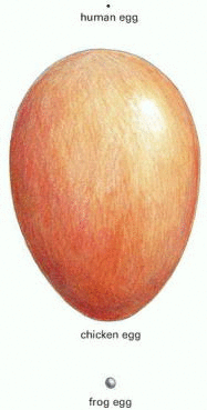
Figure 20-19
The actual sizes of three eggs. The human egg is 0.1 mm in diameter.

Figure 20-20
The relative sizes of various eggs. Sizes are compared with that of a typical somatic cell.
The egg cytoplasm contains nutritional reserves in the form of yolk, which is rich in lipids, proteins, and polysaccharides and is usually contained within discrete structures called yolk granules. In some species, each yolk granule is membrane-enclosed, whereas in others it is not. In eggs that develop into large animals outside the mother's body, yolk can account for more than 95% of the volume of the cell. In mammals, whose embryos are largely nourished by their mothers, there is little, if any, yolk.
The egg coat is another peculiarity of eggs. It is a specialized form of extracellular matrix consisting largely of glycoprotein molecules, some secreted by the egg and others deposited on it by surrounding cells. In many species, the major coat is a layer immediately surrounding the egg plasma membrane; in nonmammalian eggs, such as those of sea urchins or chickens, it is called the vitelline layer, whereas in mammalian eggs it is called the zona pellucida(Figure 20-21). This layer protects the egg from mechanical damage, and in many eggs it also acts as a species-specific barrier to sperm, admitting only those of the same or closely related species.'
https://www.ncbi.nlm.nih.gov/books/NBK26842/
' Primordial germ cells migrate to the forming gonad to become oogonia, which proliferate by mitosis for a period before differentiating into primary oocytes. At this stage (usually before birth in mammals), the first meiotic division begins: the DNA replicates so that each chromosome consists of two sister chromatids, the duplicated homologous chromosomes pair along their long axes, and crossing-over occurs between nonsister chromatids of these paired chromosomes. After these events, the cell remains arrested in prophase of division I of meiosis (in a state equivalent, as we previously pointed out, to a G2 phase of a mitotic division cycle) for a period lasting from a few days to many years, depending on the species. During this long period (or, in some cases, at the onset of sexual maturity), the primary oocytes synthesize a coat and cortical granules. In the case of large nonmammalian oocytes, they also accumulate ribosomes, yolk, glycogen, lipid, and the mRNA that will later direct the synthesis of proteins required for early embryonic growth and the unfolding of the developmental program. In many oocytes, the intensive biosynthetic activities are reflected in the structure of the chromosomes, which decondense and form lateral loops, taking on a characteristic “lampbrush” appearance, signifying that they are very busily engaged in RNA synthesis (see Figures 4-36 and 4-37).'
https://www.ncbi.nlm.nih.gov/books/NBK26842/
No comments:
Post a Comment