Buruli ulcer (/bəˈruːli/)[2] is an infectious disease characterized by the development of painless open wounds. The disease is limited to certain areas of the world, most cases occurring in Sub-Saharan Africa and Australia. The first sign of infection is a small painless nodule or area of swelling, typically on the arms or legs. The nodule grows larger over days to weeks, eventually forming an open ulcer. Deep ulcers can cause scarring of muscles and tendons, resulting in permanent disability.
Buruli ulcer is caused by skin infection with bacteria called Mycobacterium ulcerans. The mechanism by which M. ulcerans is transmitted from the environment to humans is not known, but may involve the bite of an aquatic insect or the infection of open wounds. Once in the skin, M. ulcerans grows and releases the toxin mycolactone, which blocks the normal function of cells, resulting in tissue death and immune suppression at the site of the ulcer.
The World Health Organization (WHO) recommends treating Buruli ulcer with a combination of the antibiotics rifampicin and clarithromycin. With antibiotic administration and proper wound care, small ulcers typically heal within six months. Deep ulcers and those on sensitive body sites may require surgery to remove dead tissue or repair scarred muscles or joints. Even with proper treatment, Buruli ulcer can take months to heal. Regular cleaning and dressing of wounds aids healing and prevents secondary infections.
In 2018, WHO received 2,713 reports of Buruli ulcer globally.[1] Although rare, it typically occurs in rural areas near slow-moving or stagnant water. The first written description of the disease is credited to Albert Ruskin Cook in 1897 at Mengo Hospital in Uganda. Fifty years later, the causative bacterium was isolated and identified by a group at the Alfred Hospital in Melbourne. In 1998, WHO established the Global Buruli Ulcer Initiative to coordinate global efforts to eliminate Buruli ulcer. WHO considers it a neglected tropical disease.
| Buruli ulcer | |
|---|---|
| Other names | Bairnsdale ulcer, Daintree ulcer, Mossman ulcer, Kumasi ulcer, Searls ulcer |
 | |
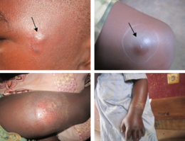
| Buruli ulcer lesions. Top-left, an early ulcer. Top-right, a larger ulcer across the lower arm and wrist. Bottom, a large ulcer on the thigh. | |
| Specialty | Infectious disease |
| Symptoms | Area of swelling that becomes an ulcer |
| Causes | Mycobacterium ulcerans |
| Treatment | Rifampicin and clarithromycin |
| Frequency | 2,713 cases reported to WHO in 2018[1] |

The first sign of Buruli ulcer is a painless swollen bump on the arm or leg, often similar in appearance to an insect bite.[1][3]Sometimes the swollen area instead appears as a patch of firm, raised skin about three centimeters across called a "plaque"; or a more widespread swelling under the skin.[1][3]
Over the course of a few weeks, the original swollen area expands to form an irregularly shaped patch of raised skin.[3][4]After about four weeks, the affected skin sloughs off leaving a painless ulcer.[1] Buruli ulcers typically have "undermined edges", the ulcer being a few centimeters wider underneath the skin than the wound itself.[4]
In some people, the ulcer may heal on its own or remain small but linger unhealed for years.[4][5] In others, it continues to grow wider and sometimes deeper, with skin at the margin dying and sloughing off. Large ulcers may extend deep into underlying tissue, causing bone infection and exposing muscle, tendon, and bone to the air.[4] When ulcers extend into muscles and tendons, parts of these tissues can be replaced by scar tissue, immobilizing the body part and resulting in permanent disability.[4] Exposed ulcers can be infected by other bacteria, causing the wound to become red, painful, and foul smelling.[6][4] Symptoms are typically limited to those caused by the wound; the disease rarely affects other parts of the body.[7]
Buruli ulcers can appear anywhere on the body, but are typically on the limbs. Ulcers are most common on the lower limbs (roughly 62% of ulcers globally) and upper limbs (24%), but can also be found on the trunk (9%), head or neck (3%), or genitals (less than 1%).[8]
The World Health Organization classifies Buruli ulcer into three categories depending on the severity of its symptoms. Category I describes a single small ulcer that is less than 5 centimetres (2.0 inches). Category II describes a larger ulcer, up to 15 centimetres (5.9 in), as well as plaques and broader swollen areas that have not yet opened into ulcers. Category III is for an ulcer larger than 15 centimeters, multiple ulcers, or ulcers that have spread to include particularly sensitive sites such as the eyes, bones, joints, or genitals.[4]
Cause[edit]
Buruli ulcer is caused by infection of the skin with the bacterium Mycobacterium ulcerans.[1] M. ulcerans is a mycobacterium, closely related to Mycobacterium marinumwhich infects aquatic animals and, rarely, humans.[9] It is more distantly related to other slow-growing mycobacteria that infect humans, such as Mycobacterium tuberculosis, which causes tuberculosis, and Mycobacterium leprae, which causes leprosy.[10] Buruli ulcer typically occurs near slow-moving or stagnant bodies of water, where M. ulcerans is found in aquatic insects, mollusks, fish, and the water itself.[11] How M. ulcerans is transmitted to humans remains unclear, but somehow bacteria enter the skin and begin to grow. Ulceration is primarily caused by the bacterial toxin mycolactone.[12] As the bacteria grow, they release mycolactone into the surrounding tissue. Mycolactone diffuses into host cells and blocks the action of Sec61, the molecular channel that serves as a gateway to the endoplasmic reticulum.[13] When Sec61 is blocked, proteins that would normally enter the endoplasmic reticulum are mistargeted to the cytosol, causing a pathological stress response that leads to cell death by apoptosis.[13] This results in tissue death at the site of infection, causing the open ulcer characteristic of the disease.[13] At the same time, Sec61 inhibition prevents cells from signaling to activate the immune system, leaving ulcers largely free of immune cells.[13] Immune cells that do reach the ulcer are killed by mycolactone, and tissue examinations of the ulcer show a core of growing bacteria surrounded by debris from dead and dying neutrophils (the most common immune cell).[14]
It is not known how M. ulcerans is introduced to humans.[1] Buruli ulcer does not spread from one person to another.[11] In areas endemic for Buruli ulcer, disease occurs near stagnant bodies of water, leading to the long-standing hypothesis that M. ulcerans is somehow transmitted to humans from aquatic environments.[15] M. ulcerans is widespread in these environments, where it can survive as free-living or in association with other aquatic organisms.[8] Live M. ulcerans has been isolated from aquatic insects, mosses, and animal feces; and its DNA has been found in water, soil, mats of bacteria and algae, fish, crayfish, aquatic insects, and other animals that live in or near water.[15] A role for biting insects in transmission has been investigated, with particular focus on mosquitoes, giant water bugs, and Naucoridae. M. ulcerans is occasionally found in these insects, and they can sometimes transmit the bacteria in laboratory settings.[8] Whether these insects are regularly involved in transmission remains unclear.[11][15]Pre-existing wounds have been implicated in disease transmission, and people who immediately wash and bandage open wounds are less likely to acquire Buruli ulcer.[16] Wearing pants and long-sleeved shirts is associated with a lower risk of Buruli ulcer, possibly by preventing insect bites or protecting wounds.[11][16]
Other ulcerative diseases can appear similar to Buruli ulcer at its various stages. The nodule that appears early in the disease can resemble a bug bite, sebaceous cyst, lipoma, onchocerciasis, other mycobacterial skin infections, or an enlarged lymph node.[4] Skin ulcers can resemble those caused by leishmaniasis, yaws, squamous cell carcinoma, Haemophilus ducreyiinfection, and tissue death due to poor circulation.[4] More diffuse lesions can resemble cellulitis and fungal infections of the skin.[4]
https://en.wikipedia.org/wiki/Buruli_ulcer
https://en.wikipedia.org/wiki/Thiotrichales
above.
No comments:
Post a Comment