Blog Archive

Tuesday, August 31, 2021

08-31-2021-1757 - bleb immune LADA T2D autoimmune

 In cell biology, a bleb is a bulge of the plasma membrane of a cell, human bioparticulate or abscess with an internal environment synonymous to that of a simple cell, characterized by a spherical, bulky morphology.[2] It is characterized by the decoupling of the cytoskeleton from the plasma membrane, degrading the internal structure of the cell, allowing the flexibility required for the cell to separate into individual bulges or pockets of the intercellular matrix.[2] Most commonly, blebs are seen in apoptosis (programmed cell death) but are also seen in other non-apoptotic functions. Blebbing, or zeiosis, is the formation of blebs.

During apoptosis, blebbing is the first phase (left) of cell disassembly.[1]

https://en.wikipedia.org/wiki/Bleb_(cell_biology)

Slowly evolving immune-mediated diabetes, or latent autoimmune diabetes in adults (LADA), is a form of diabetes that exhibits clinical features similar to both type 1 diabetes (T1D) and type 2 diabetes (T2D).[3][4]It is an autoimmune form of diabetes, similar to T1D, but patients with LADA often show insulin resistance, similar to T2D, and share some risk factors for the disease with T2D.[3] Studies have shown that LADA patients have certain types of antibodies against the insulin-producing cells, and that these cells stop producing insulin more slowly than in T1D patients.[3][5]

LADA appears to share genetic risk factors with both T1D and T2D but is genetically distinct from both.[6][7][8][9][4] Within the LADA patient group, a genetic and phenotypic heterogeneity has been observed with varying degrees of insulin resistance and autoimmunity.[5][10] With the knowledge we have today, LADA can thus be described as a hybrid form of T1D and T2D, showing phenotypic and genotypic similarities with both, as well as variation within LADA regarding the degree of autoimmunity and insulin resistance.

The concept of LADA was first introduced in 1993,[11] though The Expert Committee on the Diagnosis and Classification of Diabetes Mellitus does not recognize the term, instead including it under the standard definition of diabetes mellitus type 1.[12]

https://en.wikipedia.org/wiki/Latent_autoimmune_diabetes_in_adults

08-31-2021-1756 - 2164/5-6,7

 08-31-2021-1756 - 2164/5-6,7

08-31-2021-1755 - Lupus erythematosus autoimmune disease neurology tuberculosis leprosy immune granulocyte

Lupus erythematosus is a collection of autoimmune diseases in which the human immune systembecomes hyperactive and attacks healthy tissues.[1] Symptoms of these diseases can affect many different body systems, including jointsskinkidneysblood cellsheart, and lungs. The most common and most severe form is systemic lupus erythematosus. 

Lupus erythematosus, Atlas der Hautkrankheiten.jpg

Lupus erythematosusSpecialtyRheumatology

Cutaneous keratosis, ulcer, atrophy, and necrobiosis

https://en.wikipedia.org/wiki/Lupus_erythematosus

08-31-2021-1754 - Acute disseminated encephalomyelitis (ADEM), or acute demyelinating encephalomyelitis autoimmune disease

Acute disseminated encephalomyelitis (ADEM), or acute demyelinating encephalomyelitis, is a rare autoimmune disease marked by a sudden, widespread attack of inflammation in the brain and spinal cord. As well as causing the brain and spinal cord to become inflamed, ADEM also attacks the nerves of the central nervous system and damages their myelin insulation, which, as a result, destroys the white matter. It is often triggered by a viral infection or (very rarely) vaccinations.[1][2][3][4][5][6]

ADEM's symptoms resemble the symptoms of multiple sclerosis (MS), so the disease itself is sorted into the classification of the multiple sclerosis borderline diseases. However, ADEM has several features that distinguish it from MS.[7] Unlike MS, ADEM occurs usually in children and is marked with rapid fever, although adolescents and adults can get the disease too. ADEM consists of a single flare-up whereas MS is marked with several flare-ups (or relapses), over a long period of time. Relapses following ADEM are reported in up to a quarter of patients, but the majority of these 'multiphasic' presentations following ADEM likely represent MS.[8] ADEM is also distinguished by a loss of consciousness, coma and death, which is very rare in MS, except in severe cases.

It affects about 8 per 1,000,000 people per year.[9] Although it occurs in all ages, most reported cases are in children and adolescents, with the average age around 5 to 8 years old.[10][11][12][13] The disease affects males and females almost equally.[14] ADEM shows seasonal variation with higher incidence in winter and spring months which may coincide with higher viral infections during these months.[13] The mortality rate may be as high as 5%; however, full recovery is seen in 50 to 75% of cases with increase in survival rates up to 70 to 90% with figures including minor residual disability as well.[15] The average time to recover from ADEM flare-ups is one to six months.

ADEM produces multiple inflammatory lesions in the brain and spinal cord, particularly in the white matter. Usually these are found in the subcortical and central white matter and cortical gray-white junction of both cerebral hemispherescerebellumbrainstem, and spinal cord,[16] but periventricular white matter and gray matter of the cortexthalami and basal ganglia may also be involved.

When a person has more than one demyelinating episode of ADEM, the disease is then called recurrent disseminated encephalomyelitis[17] or multiphasic disseminated encephalomyelitis[18] (MDEM). Also, a fulminant course in adults has been described.[19] 

https://en.wikipedia.org/wiki/Acute_disseminated_encephalomyelitis

08-31-2021-1753 - Antiphospholipid syndrome, or antiphospholipid antibody syndrome (APS or APLS), phospholipid lipid lipopolysaccharide LPS petersen autoimmune hypercoagulable

Antiphospholipid syndrome, or antiphospholipid antibody syndrome (APS or APLS), is an autoimmunehypercoagulable state caused by antiphospholipid antibodies. APS provokes blood clots (thrombosis) in both arteries and veins as well as pregnancy-related complications such as miscarriagestillbirthpreterm delivery, and severe preeclampsia. Although the exact etiology of APS is still not clear, genetics is believed to play a key role in the development of the disease.[2] The diagnostic criteria require one clinical event (i.e. thrombosis or pregnancy complication) and two positive blood test results spaced at least three months apart that detect lupus anticoagulantanti-apolipoprotein antibodies, or anti-cardiolipin antibodies.[3]

Antiphospholipid syndrome can be primary or secondary. Primary antiphospholipid syndrome occurs in the absence of any other related disease. Secondary antiphospholipid syndrome occurs with other autoimmune diseases, such as systemic lupus erythematosus (SLE). In rare cases, APS leads to rapid organ failure due to generalised thrombosis; this is termed "catastrophic antiphospholipid syndrome" (CAPS or Asherson syndrome) and is associated with a high risk of death.

Antiphospholipid syndrome often requires treatment with anticoagulant medication such as heparin to reduce the risk of further episodes of thrombosis and improve the prognosis of pregnancy. Warfarin(brand name Coumadin) is not used during pregnancy because it can cross the placenta, unlike heparin, and is teratogenic. 

https://en.wikipedia.org/wiki/Antiphospholipid_syndrome

08-31-2021-1752 - Autoimmune autonomic ganglionopathy (AAG) immune AChR acetylcholinesterase inhibitor antibody autoimmune state (reversible) disease (irreversible) [with exp and control birth to death lab]

Autoimmune autonomic ganglionopathy (AAG) is a rare form of dysautonomia in which the patient’s immune system produces ganglionic anti-nicotinic acetylcholine receptor (AChR) antibodies, inhibiting ganglionic AChR currents and impairing transmission in autonomic ganglia.[1] Symptoms onset can be acute, subacute or gradual. 

https://en.wikipedia.org/wiki/Autoimmune_autonomic_ganglionopathy

08-31-2021-1749 - Neuropsychiatric systemic lupus erythematosus or NPSLE lupus autoimmune disease immunology immune NAT nat neurocystcercosis neurosarcodoisis Neurosarcoidosis

 Neuropsychiatric systemic lupus erythematosus or NPSLE refers to the neurological and psychiatricmanifestations of systemic lupus erythematosus. SLE is a disease in which the immune system attacks the body's own cells and tissues. It can affect various organs or systems of the body. It is estimated that over half of people with SLE have neuropsychiatric involvement.[1]

https://en.wikipedia.org/wiki/Neuropsychiatric_systemic_lupus_erythematosus

Neuropsychiatric systemic lupus erythematosus
Other namesCNS lupus, lupus cerebritis
SpecialtyRheumatologyNeurology

The American College of Rheumatology (ACR) has outlined 19 syndromes that are seen in NPSLE. These syndromes encompass disorders of the centraland peripheral nervous systems:[2]

Each of the 19 syndromes are also stand-alone diagnoses, which can occur with or without lupus.

The majority of cases involve the central nervous system (CNS), which consists of the brain and spinal cord.[2] The most common CNS syndromes are headache and mood disorder.[1]

Though neuropsychiatric lupus is sometimes referred to as "CNS lupus", it can also affect the peripheral nervous system (PNS). Between 10-15% of people with NPSLE have PNS involvement.[3] Mononeuropathy and polyneuropathy are the most common PNS syndromes.[1]

Other syndromes[edit]

Some neurological syndromes outside of the ACR classification may also be considered NPSLE manifestations. These include neuromyelitis opticaposterior reversible encephalopathy syndromesmall fiber neuropathy,[4] and Lambert–Eaton myasthenic syndrome.[5]


08-31-2021-1748 - Nuclear bodies (also known as nuclear domains, or nuclear dots)

 Nuclear bodies (also known as nuclear domains, or nuclear dots) are membraneless structures found in the cell nuclei of eukaryotic cells.[1] Nuclear bodies include Cajal bodies, the nucleolus, and promyelocytic leukemia protein (PML) nuclear bodies (also called PML oncogenic dots).[2] Nuclear bodies also include ND10s. ND stands for nuclear domain, and 10 refers to the number of dots seen.[3]

Nuclear bodies were first seen as prominent interchromatin structures in the nuclei of malignant or hyperstimulated animal cells[4][5] identified using anti-sp100 autoantibodies from primary biliary cirrhosis and subsequently the promyelocytic leukemia (PML) factor, but appear also to be elevated in many autoimmune and cancerous diseases.[6] Nuclear dots are metabolically stable and resistant to nuclease digestion and salt extraction.[7]

A nuclear body subtype is a clastosome suggested to be a site of protein degradation.[8]

https://en.wikipedia.org/wiki/Nuclear_bodies

08-31-2021-1748 - Autoimmune encephalitis

Autoimmune encephalitis

 https://en.wikipedia.org/wiki/Autoimmune_encephalitis


08-31-2021-1748 - Autoimmune hemolytic anemia (AIHA)

Autoimmune hemolytic anemia (AIHA) occurs when antibodies directed against the person's own red blood cells (RBCs) cause them to burst (lyse), leading to an insufficient number of oxygen-carrying red blood cells in the circulation. The lifetime of the RBCs is reduced from the normal 100–120 days to just a few days in serious cases.[1][2] The intracellular components of the RBCs are released into the circulating blood and into tissues, leading to some of the characteristic symptoms of this condition. The antibodies are usually directed against high-incidence antigens, therefore they also commonly act on allogenic RBCs (RBCs originating from outside the person themselves, e.g. in the case of a blood transfusion).[3] AIHA is a relatively rare condition, affecting one to three people per 100,000 per year.[4] Autoimmune hemolysis might be a precursor of later onset systemic lupus erythematosus.[5]

The terminology used in this disease is somewhat ambiguous. Although MeSH uses the term "autoimmune hemolytic anemia",[6] some sources prefer the term "immunohemolytic anemia" so drug reactions can be included in this category.[7][8] The National Cancer Institute considers "immunohemolytic anemia", "autoimmune hemolytic anemia", and "immune complex hemolytic anemia" to all be synonyms.[9] 

https://en.wikipedia.org/wiki/Autoimmune_hemolytic_anemia

08-31-2021-1747 - Autoimmune retinopathy (AIR) Grande

Autoimmune retinopathy (AIR) is a rare disease in which the patient's immune system attacks proteins in the retina, leading to loss of eyesight. The disease is poorly understood, but may be the result of cancer or cancer chemotherapy.[1] The disease is an autoimmune condition characterized by vision loss, blind spots, and visual field abnormalities. It can be divided into cancer-associated retinopathy (CAR) and melanoma-associated retinopathy (MAR).[2] The condition is associated with retinal degeneration caused by autoimmune antibodies recognizing retinal proteins as antigens and targeting them.[3] AIR's prevalence is extremely rare, with CAR being more common than MAR.[2] It is more commonly diagnosed in females (approximately 60% of diagnosed patients are females) in the age range of 50-60.[2]

https://en.wikipedia.org/wiki/Autoimmune_retinopathy

08-31-2021-1747 - Granuloma annulare

Granuloma annulare (GA) is a common, sometimes chronic skin condition which presents as reddish bumps on the skin arranged in a circle or ring.[1] It can initially occur at any age, though two-thirds of patients are under 30 years old, and it is seen most often in children and young adults. Females are two times as likely to have it than males.[2]

https://en.wikipedia.org/wiki/Granuloma_annulare

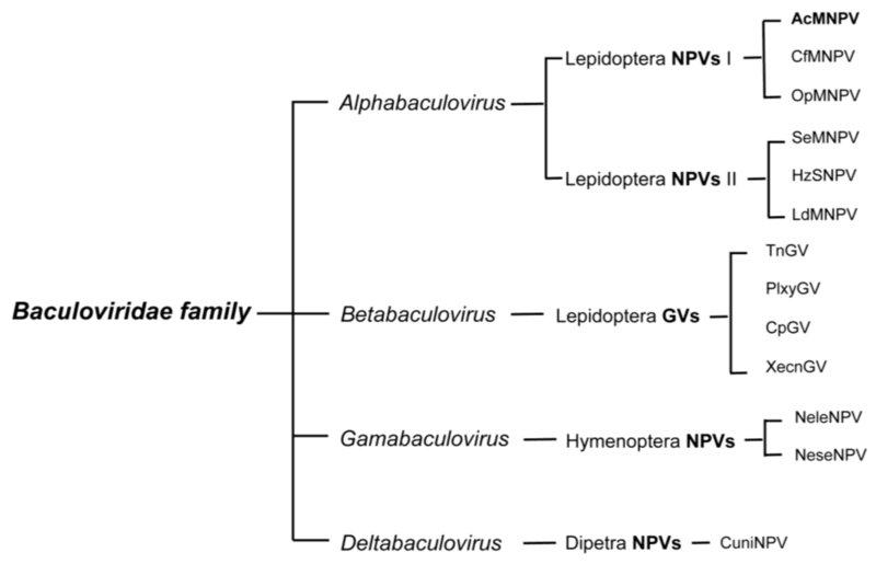
08-31-2021-1637 - deltabaculovirus - Betabaculovirus Unassigned dsDNA deltabaculovirus NPV dipetra hymenoptera diptera lepidoptera gammabaculovirus gamma alpha beta delta

Original file (1,024 × 659 pixels, file size: 51 KB, MIME type: image/png) 

https://microbewiki.kenyon.edu/index.php/File:Baculovirus_taxonomy.png


Domain: Viruses

Group: dsDNA

Order: Unassigned

Family Baculoviridae

Genus: Betabaculovirus

Type Species

Cydia pomonella granulovirus

https://microbewiki.kenyon.edu/index.php/Granulosis_Virus


Cydia pomonella granulovirus

Poor Unfortunate Souls - The Little Mermaid (1989)


08312021-1625

The Little Mermaid On Broadway - Part Of Your World


083120211600

08-31-2021-1151 - Sea god's palace messengers; legend

Legend has it that they beach themselves before underwater earthquakes or tsunamis.

Known in Japan as a “messenger from the sea god’s palace”, oarfish are thought to live around a kilometre underwater and surface when sick or dying.

https://www.independent.co.uk/news/world/asia/japan-oarfish-washed-earthquake-tsunami-legend-a8759846.html