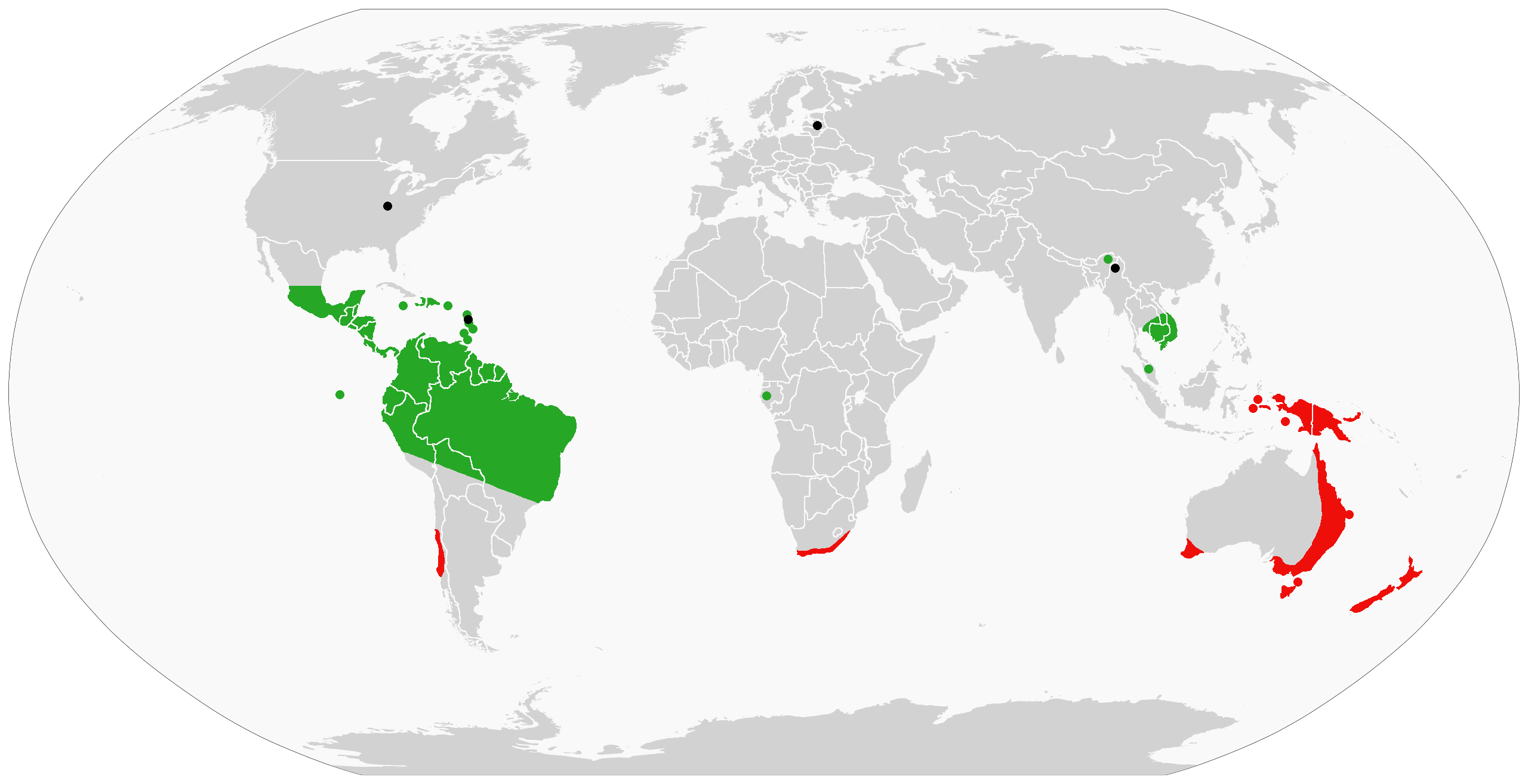
An amebocyte or amoebocyte (/əˈmiː.bə.saɪt/) is a mobile cell (moving like an amoeba) in the body of invertebrates including echinoderms, molluscs, tunicates, sponges and some chelicerates. They move by pseudopodia. Similarly to some of the white blood cells of vertebrates, in many species amebocytes are found in the blood or body fluid and play a role in the defense of the organism against pathogens. Depending on the species, an amebocyte may also digest and distribute food, dispose of wastes, form skeletal fibers, fight infections, and change into other cell types.
Limulus amebocyte lysate, an aqueous extract of amebocytes from the Atlantic horseshoe crab (Limulus polyphemus), is commonly used in a test to detect bacterial endotoxins.[1]
In sponges, amebocytes, also known as archaeocytes, are cells found in the mesohyl that can transform into any of the animal's more specialized cell types.[2][3]
In tunicates they are blood cells and use pseudopodia to attack pathogens that enter the blood, transport nutrients, get rid of waste products, and grow/repair the tunica.[4] In older literature, the term amebocyte is sometimes used as a synonym of phagocyte.
References[edit]
- ^ Levin J, Bang F.B. (1968). "Clottable Protein in Limulus: Its Localization and Kinetics of Its Coagulation by Endotoxin". Thromb. Diath. Haemorrh. 19 (1): 186–97. PMID 5690028.
- ^ "An Online Introduction to the Biology of Animals and Plants - Sponges and Cnidaria".
- ^ "The Porifera - Invertebrate Biology Course".
- ^ Cima, Francesca; Ballarin, Loriano; Gasparini, Fabio; Burighel, Paolo (2006-01-01). "External amebocytes guard the pharynx entry in a tunicate (Ascidiacea)". Developmental & Comparative Immunology. 30 (5): 463–472. doi:10.1016/j.dci.2005.07.004. PMID 16182366.

The body cavity is known as a "pseudocoel", or haemocoel. Unlike a true coelom, a pseudocoel is not fully enclosed by a cell layer derived from the embryonic mesoderm. A coelom is, however, formed around the gonads and the waste-eliminating nephridia.[8] As the name haemocoel suggests, the body cavity is filled with a blood-like liquid in which all the organs are embedded; in this way, they can be easily supplied with nutrients circulating in the blood. This liquid is colourless as it does not contain pigments; for this reason, it serves only a limited role in oxygen transport. Two different types of blood cells (or haemocytes) circulate in the fluid: amoebocytes and nephrocytes. The amoebocytes probably function in protection from bacteria and other foreign bodies; in some species, they also play a role in reproduction. Nephrocytes absorb toxins or convert them into a form suitable for elimination by the nephridia.[citation needed] The haemocoel is divided by a horizontal partition, the diaphragm, into two parts: the pericardial sinus along the back and the perivisceral sinus along the belly. The former encloses the tube-like heart, and the latter, the other organs. The diaphragm is perforated in many places, enabling the exchange of fluids between the two cavities.[citation needed] The heart itself is a tube of annular muscles consisting of epithelial tissues, with two lateral openings (ostia) per segment. While it is not known whether the rear end is open or closed, from the front, it opens directly into the body cavity. Since there are no blood vessels, apart from the fine vessels running between the muscle layers of the body wall and a pair of arteries that supply the antennae, this is referred to as an open circulation.[citation needed] The timing of the pumping procedure can be divided into two parts: diastole and systole. During diastole, blood flows through the ostia from the pericardial sinus (the cavity containing the heart) into the heart. When the systole begins, the ostia close and the heart muscles contract inwards, reducing the volume of the heart. This pumps the blood from the front end of the heart into the perivisceral sinus containing the organs. In this way, the various organs are supplied with nutrients before the blood finally returns to the pericardial sinus via the perforations in the diaphragm. In addition to the pumping action of the heart, body movements also have an influence on circulation.[citation needed]
Respiration[edit]
Oxygen uptake occurs to an extent via simple diffusion through the entire body surface, with the coxal vesicles on the legs possibly being involved in some species. However, of most importance is gas exchange via fine unbranched tubes, the tracheae, which draw oxygen from the surface deep into the various organs, particularly the heart. The walls of these structures, which are less than three micrometers thick in their entirety, consist only of an extremely thin membrane through which oxygen can easily diffuse. The tracheae originate at tiny openings, the spiracles, which themselves are clustered together in dent-like recesses of the outer skin, the atria. The number of "tracheae bundles" thus formed is on average around 75 per body segment; they accumulate most densely on the back of the organism.[citation needed] Unlike the arthropods, the velvet worms are unable to control the openings of their tracheae; the tracheae are always open, entailing considerable water loss in arid conditions. Water is lost twice as fast as in earthworms and forty times faster than in caterpillars.[21] For this reason, velvet worms are dependent upon habitats with high air humidity.[citation needed]
No comments:
Post a Comment