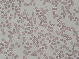
Spherocytosis is the presence in the blood of spherocytes, i.e erythrocytes (red blood cells) that are sphere-shaped rather than bi-concave disk shaped as normal. Spherocytes are found in all hemolytic anemias to some degree. Hereditary spherocytosis and autoimmune hemolytic anemiaare characterized by having only spherocytes.[1]
| Spherocytosis | |
|---|---|
 | |
| Spherocytosis seen in a peripheral blood smear from a patient with hereditary spherocytosis | |
| Specialty | Hematology |
Spherocytes are found in immunologically-mediated hemolytic anemias and in hereditary spherocytosis, but the former would have a positive direct Coombs test and the latter would not. The misshapen but otherwise healthy red blood cells are mistaken by the spleen for old or damaged red blood cells and it thus constantly breaks them down, causing a cycle whereby the body destroys its own blood supply (auto-hemolysis). A complete blood count (CBC) may show increased reticulocytes, a sign of increased red blood cell production, and decreased hemoglobin and hematocrit. The term "non-hereditary spherocytosis" is occasionally used, albeit rarely.[2]
Lists of causes:[3]
- Warm autoimmune hemolytic anemia
- Cold autoimmune hemolytic anemia/paroxysmal cold hemoglobinuria
- Acute and delayed hemolytic transfusion reactions
- ABO hemolytic diseases of newborn/Rh hemolytic disease of newborn
- Hereditary spherocytosis
- Intravenous water infusion or drowning (fresh water)
- Hypophosphatemia
- Bartonellosis
- Snake bites
- Hyposplenism
- Rh-null phenotype
Spherocytosis most often refers to hereditary spherocytosis. This is caused by a molecular defect in one or more of the proteins of the red blood cell cytoskeleton, including spectrin, ankyrin, Band 3, or Protein 4.2. Because the cell skeleton has a defect, the blood cell contracts to a sphere, which is its most surface tension efficient and least flexible configuration. Though the spherocytes have a smaller surface area through which oxygen and carbon dioxide can be exchanged, they in themselves perform adequately to maintain healthy oxygen supplies. However, they have a high osmoticfragility—when placed into water, they are more likely to burst than normal red blood cells. These cells are more prone to physical degradation.[citation needed]
In short, spherocytosis has an attribute of decreased cell deformability.[4]
Spherocytosis can be diagnosed in Peripheral blood film by seeing spherical red blood cells rather than biconcave. Because spherical red blood cells are more prone to lysis in water (because they lack some proteins in their cytoskeleton) there will be increased osmotic fragility on acidified glycerol lysis test.[citation needed]
See also[edit]
https://en.wikipedia.org/wiki/Spherocytosis
https://nikiyaantonbettey.blogspot.com/2021/08/08-28-2021-2312-ankyrin-repeat.html
No comments:
Post a Comment