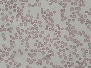
Hereditary spherocytosis is an abnormality of red blood cells, or erythrocytes. It is a chronic disease with no cure. The disorder is caused by mutations in genes relating to membrane proteins that allow for the erythrocytes to change shape. The abnormal erythrocytes are sphere-shaped (spherocytosis) rather than the normal biconcave disk shaped. Dysfunctional membrane proteins interfere with the cell's ability to be flexible to travel from the arteries to the smaller capillaries. This difference in shape also makes the red blood cells more prone to rupture.[1] Cells with these dysfunctional proteins are degraded in the spleen. This shortage of erythrocytes results in hemolytic anemia.
It was first described in 1871. It is the most common cause of inherited hemolysis in European and North American Caucasian populations, with an incidence of 1 in 5000 births. The clinical severity of HS varies from symptom-free carrier to severe hemolysis because the disorder exhibits incomplete penetrance in its expression.
Symptoms include anemia, jaundice, splenomegaly, and fatigue.[2] Furthermore, the detritus of the broken-down blood cells – unconjugated or indirect bilirubin – accumulates in the gallbladder, and can cause pigmented gallstones to develop. In chronic patients, an infection or other illness can cause an increase in the destruction of red blood cells, resulting in the appearance of acute symptoms, a hemolytic crisis. On a blood smear, Howell-Jolly bodies may be seen within red blood cells. Primary treatment for patients with symptomatic HS has been total splenectomy, which eliminates the hemolytic process, allowing normal hemoglobin, reticulocyte and bilirubin levels. Spherocytosis patients who are heterozygous for a hemochromatosis gene may suffer from iron overload, despite the hemochromatosis genes being recessive.[3][4]
Acute cases can threaten to cause hypoxia through anemia and acute kernicterus through high blood levels of bilirubin, particularly in newborns. Most cases can be detected soon after birth. An adult with this disease should have their children tested, although the presence of the disease in children is usually noticed soon after birth. Occasionally, the disease will go unnoticed until the child is about 4 or 5 years of age. A person may also be a carrier of the disease and show no signs or symptoms of the disease. Other symptoms may include abdominal pain that could lead to the removal of the spleen and/or gallbladder.
| Hereditary spherocytosis | |
|---|---|
| Other names | Minkowski–Chauffard syndrome |
 | |
| Peripheral blood smear from patient with hereditary spherocytosis | |
| Specialty | Hematology |
https://en.wikipedia.org/wiki/Hereditary_spherocytosis
Kernicterus is a bilirubin-induced brain dysfunction.[1] The term was coined in 1904 by Schmorl. Bilirubin is a naturally occurring substance in the body of humans and many other animals, but it is neurotoxic when its concentration in the blood is too high, a condition known as hyperbilirubinemia. Hyperbilirubinemia may cause bilirubin to accumulate in the grey matter of the central nervous system, potentially causing irreversible neurological damage. Depending on the level of exposure, the effects range from clinically unnoticeable to severe brain damage and even death.
When hyperbilirubinemia increases past a mild level, it leads to jaundice, raising the risk of progressing to kernicterus. When this happens in adults, it is usually because of liver problems. Newborns are especially vulnerable to hyperbilirubinemia-induced neurological damage, because in the earliest days of life, the still-developing liver is heavily exercised by the breakdown of fetal hemoglobin as it is replaced with adult hemoglobin and the blood–brain barrier is not as developed. Mildly elevated serum bilirubin levels are common in newborns, and neonatal jaundiceis not unusual, but bilirubin levels must be carefully monitored in case they start to climb, in which case more aggressive therapy is needed, usually via light therapy but sometimes even via exchange transfusion.
| Kernicterus | |
|---|---|
 | |
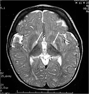
| Brain MRI. Hyperintense basal ganglia lesions on T2-weighted images. | |
| Specialty | Psychiatry, Neurology, Pediatrics |
| Diagnostic method | physical examination of moro reflex |
https://en.wikipedia.org/wiki/Kernicterus
In short, spherocytosis has an attribute of decreased cell deformability.[4]
https://en.wikipedia.org/wiki/Spherocytosis
Hypersensitivity pneumonitis (HP) or extrinsic allergic alveolitis (EAA) is a rare immune system disorder that affects the lungs.[1] It is an inflammation of the airspaces (alveoli) and small airways (bronchioles) within the lung, caused by hypersensitivity to inhaled organic dusts and molds.[2] People affected by this type of lung inflammation (pneumonitis) are commonly exposed to the dust and mold by their occupation or hobbies.[2]
https://en.wikipedia.org/wiki/Hypersensitivity_pneumonitis
No comments:
Post a Comment