United Nations (1972). Biological Weapons Convention.
While the history of biological weapons use goes back more than six centuries to the siege of Caffa in 1346,[8] international restrictions on biological weapons began only with the 1925 Geneva Protocol, which prohibits the use but not the possession or development of chemical and biological weapons.[9] Upon ratification of the Geneva Protocol, several countries made reservations regarding its applicability and use in retaliation.[10] Due to these reservations, it was in practice a "no-first-use" agreement only.[11]
The 1972 Biological Weapons Convention (BWC) supplements the Geneva Protocol by prohibiting the development, production, acquisition, transfer, stockpiling and use of biological weapons.[12] Having entered into force on 26 March 1975, the BWC was the first multilateral disarmament treaty to ban the production of an entire category of weapons of mass destruction.[12] As of March 2021, 183 states have become party to the treaty.[2] The BWC is considered to have established a strong global norm against biological weapons,[13] which is reflected in the treaty’s preamble, stating that the use of biological weapons would be "repugnant to the conscience of mankind".[14] However, the BWC’s effectiveness has been limited due to insufficient institutional support and the absence of any formal verification regime to monitor compliance.[15]
In 1985, the Australia Group was established, a multilateral export control regime of 43 countries aiming to prevent the proliferation of chemical and biological weapons.[16]
In 2004, the United Nations Security Council passed Resolution 1540, which obligates all UN Member States to develop and enforce appropriate legal and regulatory measures against the proliferation of chemical, biological, radiological, and nuclear weapons and their means of delivery, in particular, to prevent the spread of weapons of mass destruction to non-state actors.[17]
Rudimentary forms of biological warfare have been practiced since antiquity.[12] The earliest documented incident of the intention to use biological weapons is recorded in Hittite texts of 1500–1200 BCE, in which victims of tularemia were driven into enemy lands, causing an epidemic.[13] The Assyrians poisoned enemy wells with the fungus ergot, though with unknown results. Scythian archers dipped their arrows and Roman soldiers their swords into excrements and cadavers – victims were commonly infected by tetanus as result.[14] In 1346, the bodies of Mongol warriors of the Golden Horde who had died of plague were thrown over the walls of the besieged Crimean city of Kaffa. Specialists disagree about whether this operation was responsible for the spread of the Black Death into Europe, Near East and North Africa, resulting in the deaths of approximately 25 million Europeans.[15][16][17][18]
Biologicals were extensively used in many parts of Africa from the sixteenth century AD, most of the time in the form of poisoned arrows, or powder spread on the war front as well as poisoning of horses and water supply of the enemy forces.[19][20] In Borgu, there were specific mixtures to kill, hypnotize, make the enemy bold, and to act as an antidote against the poison of the enemy as well. The creation of biologicals was reserved for a specific and professional class of medicine-men.[20]
Anti-crop/anti-vegetation/anti-fisheries[edit]
The United States developed an anti-crop capability during the Cold War that used plant diseases (bioherbicides, or mycoherbicides) for destroying enemy agriculture. Biological weapons also target fisheries as well as water-based vegetation. It was believed that the destruction of enemy agriculture on a strategic scale could thwart Sino-Soviet aggression in a general war. Diseases such as wheat blast and rice blast were weaponized in aerial spray tanks and cluster bombs for delivery to enemy watersheds in agricultural regions to initiate epiphytotic (epidemics among plants). On the other hand, some sources report that these agents were stockpiled but never weaponized.[85] When the United States renounced its offensive biological warfare program in 1969 and 1970, the vast majority of its biological arsenal was composed of these plant diseases.[86] Enterotoxins and Mycotoxins were not affected by Nixon's order.
Though herbicides are chemicals, they are often grouped with biological warfare and chemical warfare because they may work in a similar manner as biotoxins or bioregulators. The Army Biological Laboratory tested each agent and the Army's Technical Escort Unit was responsible for the transport of all chemical, biological, radiological (nuclear) materials.
Biological warfare can also specifically target plants to destroy crops or defoliate vegetation. The United States and Britain discovered plant growth regulators (i.e., herbicides) during the Second World War, which were then used by the UK in the counterinsurgency operations of the Malayan Emergency. Inspired by the use in Malaysia, the US military effort in the Vietnam War included a mass dispersal of a variety of herbicides, famously Agent Orange, with the aim of destroying farmland and defoliating forests used as cover by the Viet Cong.[87] Sri Lanka deployed military defoliants in its prosecution of the Eelam War against Tamil insurgents.[88]
Anti-livestock[edit]
During World War I, German saboteurs used anthrax and glanders to sicken cavalry horses in U.S. and France, sheep in Romania, and livestock in Argentina intended for the Entente forces.[89] One of these German saboteurs was Anton Dilger. Also, Germany itself became a victim of similar attacks – horses bound for Germany were infected with Burkholderia by French operatives in Switzerland.[90]
During World War II, the U.S. and Canada secretly investigated the use of rinderpest, a highly lethal disease of cattle, as a bioweapon.[89][91]
In the 1980s Soviet Ministry of Agriculture had successfully developed variants of foot-and-mouth disease, and rinderpest against cows, African swine fever for pigs, and psittacosis to kill the chicken. These agents were prepared to spray them down from tanks attached to airplanes over hundreds of miles. The secret program was code-named "Ecology".[52]
During the Mau Mau Uprising in 1952, the poisonous latex of the African milk bush was used to kill cattle.[92]
See also[edit]
Above. CL1 civilian applicants and subjects various stories.
See also[edit]
- Animal-borne bomb attacks
- Antibiotic resistance
- Asymmetric warfare
- Baker Island
- Bioaerosol
- Biological contamination
- Biological pest control
- Biosecurity
- Chemical weapon
- Counterinsurgency
- Discredited AIDS origins theories
- Enterotoxin
- Entomological warfare
- Ethnic bioweapon
- Herbicidal warfare
- Human experimentation in the United States
- John W. Powell
- Johnston Atoll Chemical Agent Disposal System
- List of CBRN warfare forces
- Plum Island Animal Disease Center
- Project 112
- Project AGILE
- Project SHAD
- McNeill's law
- Military Animal
- Animal as weapon
- Mycotoxin
- Rhodesia and weapons of mass destruction
- Ten Threats
- Trichothecene
- Yellow rain
- Vaccines
https://en.wikipedia.org/wiki/Biological_warfare
| When Dee Dee hides the core of Dexter's nuclear power lamp, he must decipher her clues within one hour to find it. | |||||||
| 23c | 10c | "Germ Warfare" | Robert Alvarez and Genndy Tartakovsky | Zeke Kamm | Ace Conrad and Genndy Tartakovsky | September 17, 1997 | 208c (34-5434) |
|---|---|---|---|---|---|---|---|
| When his family is suffering from the flu, Dexter avoids contracting the illness, but his efforts are thwarted by a sickly Dee Dee. | |||||||
"Germ Warfare" was the 11th episode of the first season of the TV series M*A*S*H. It originally aired on December 10, 1972.
Guest cast is Patrick Adiarte as Ho-Jon, Timothy Brown as Spearchucker Jones, Byron Chung as P.O.W., Odessa Cleveland as Ginger, Bob Gooden as Boone, and Karen Philipp as Lt. Dish.[1][2][3][4][5][6] The episode features the final appearances in the series of Captain Jones, Lt. Dish and Pvt. Boone—all simply disappear after this point, without comment or explanation.
https://en.wikipedia.org/wiki/Germ_Warfare_(M*A*S*H)
Simulants[edit]
Simulants are organisms or substances which mimic physical or biological properties of real biological agents, without being pathogenic. They are used to study the efficiency of various dissemination techniques or the risks caused by the use of biological agents in bioterrorism.[6] To simulate dispersal, attachment or the penetration depth in human or animal lungs, simulants must have particle sizes, specific weight and surface properties, similar to the simulated biological agent.
The typical size of simulants (1–5 µm) enables it to enter buildings with closed windows and doors and penetrate deep into the lungs. This bears a significant health risk, even if the biological agent is normally not pathogenic.
- Bacillus globigii (historically named Bacillus subtilis in the context of bio-agent simulants) (BG, BS, or U)
- Serratia marcescens (SM or P)
- Aspergillus fumigatus mutant C-2 (AF)
- Escherichia coli (EC)
- Bacillus thuringiensis (BT)
- Erwinia herbicola (current accepted name: Pantoea agglomerans) (EH)
- Fluorescent particles such as Zinc cadmium sulfide, ZnCdS (FP)
https://en.wikipedia.org/wiki/Biological_agent
The gregarines are a group of Apicomplexan alveolates, classified as the Gregarinasina[1] or Gregarinia. The large (roughly half a millimeter) parasites inhabit the intestines of many invertebrates. They are not found in any vertebrates. However, gregarines are closely related to both Toxoplasma and Plasmodium, which cause toxoplasmosis and malaria, respectively. Both protists use protein complexes similar to those that are formed by the gregarines for gliding motility and invading target cells.[2][3] This makes them excellent models for studying gliding motility with the goal of developing treatment options for toxoplasmosis and malaria. Thousands of different species of gregarines are expected to be found in insects, and 99% of these gregarines still need to be described. Each insect can be the host of multiple species.[4][5] One of the most studied gregarines is Gregarina garnhami. In general, gregarines are regarded as very successful parasites, as their hosts are spread over the entire planet.[6]
| Gregarinasina | |
|---|---|
 | |
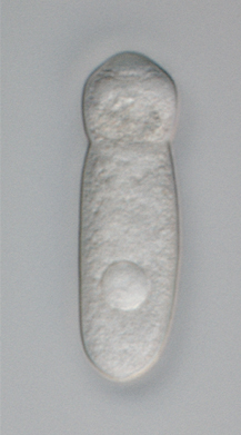
| A live specimen of a septate (or cephaline) gregarine showing the distinctive "head"-like section of the trophozoite containing the epimerite at its anterior end. Septate gregarines are intestinal parasites of arthropods. | |
| Scientific classification | |
| Domain: | |
| (unranked): | |
| (unranked): | |
| Phylum: | |
| Class: | |
| Subclass: | Gregarinasina |
| Orders | |
| Synonyms | |
| |
Gregarines occur in both aquatic and terrestrial environments. Although they are usually transmitted by the orofaecal route, some are transmitted with the host's gametes during copulation (Monocystis).
In all species, four or more sporozoites (the precise number depends on the species) equipped with an apical complex escape from the oocysts, a process called excystation, find their way to the appropriate body cavity, and penetrate host cells in their immediate environment. The sporozoites emerge within the host cell, begin to feed, and develop into larger trophozoites. In some species, the sporozoites and trophozoites are capable of asexual replication — a process called schizogony or merogony. Most species, however, appear to lack schizogony in their lifecycles.
In all species, two mature trophozoites eventually pair up in a process known as syzygy and develop into gamonts. During syzygy, gamont orientation differs between species (side-to-side, head-to-tail). A gametocyst wall forms around each pair of gamonts, which then begin to divide into hundreds of gametes. Zygotes are produced by the fusion of two gametes, and these, in turn, become surrounded by an oocyst wall. Within the oocyst, meiosisoccurs, yielding the sporozoites. Hundreds of oocysts accumulate within each gametocyst and these are released via host's faeces or via host death and decay.
Gregarines have been reported to infect over 3000 invertebrate species.[7]
- Meiosis occurs in all species.
- Monoxenous — only one host occurs in lifecycle for almost all species.
- Mitochondria have tubular cristae and are often distributed near the cell periphery.
- Apical complex occurs in the sporozoite stage, but is lost in the trophozoite stage in eugregarines and neogregarines.
- Trophozoites have a large and conspicuous nucleus and nucleolus.
- They inhabit extracellular body cavities of invertebrates such as the intestines, coeloms, and reproductive vesicles.
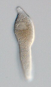
- Attachment to host occurs by a mucron (aseptate gregarines) or an epimerite (septate gregarines); some gregarines (urosporidians) float freely within extracellular body cavities (coelom).
The parasites are relatively large, spindle-shaped cells, compared to other apicomplexans and eukaryotes in general (some species are > 850 µm in length). Most gregarines have longitudinal epicytic folds (bundles of microtubules beneath the cell surface with nematode like bending behaviour): crenulations are instead found in the urosporidians.
Molecular biology[edit]
The gregarines are able to move and change direction along a surface through gliding motility without the use of cilia, flagella, or lamellipodia.[14] This is accomplished through the use of an actin and myosin complex.[15] The complexes require an actin cytoskeleton to perform their gliding motions.[16] In the proposed ‘capping’ model, an uncharacterized protein complex moves rearward, moving the parasites forward.[17]
The gregarines are among the oldest known parasites, having been described by the physician Francesco Redi in 1684.[18]
The first formal description was made by Dufour in 1828. He created the genus Gregarina and described Gregarina ovata from Folficula aricularia. He considered them to be parasitic worms. Koelliker recognised them as protozoa in 1848.
https://en.wikipedia.org/wiki/Gregarinasina
https://www.osha.gov/biological-agents


No comments:
Post a Comment