For the organelle called conoid used by intracellular parasites, see myzocytosis.
In geometry a conoid (Greek: κωνος cone and -ειδης similar) is a ruled surface, whose rulings (lines) fulfill the additional conditions
- (1) All rulings are parallel to a plane, the directrix plane.
- (2) All rulings intersect a fixed line, the axis.
- The conoid is a right conoid, if its axis is perpendicular to its directrix plane. Hence all rulings are perpendicular to the axis.
Because of (1) any conoid is a Catalan surface and can be represented parametrically by
Any curve with fixed parameter is a ruling, describes the directrix and the vectors are all parallel to the directrix plane. The planarity of the vectors can be represented by
- .
- If the directrix is a circle the conoid is called circular conoid.
The term conoid was already used by Archimedes in his treatise On conoids and spheroides.
https://en.wikipedia.org/wiki/Conoid
In geometry, a right conoid is a ruled surface generated by a family of straight lines that all intersect perpendicularly to a fixed straight line, called the axis of the right conoid.
Using a Cartesian coordinate system in three-dimensional space, if we take the z-axis to be the axis of a right conoid, then the right conoid can be represented by the parametric equations:
where h(u) is some function for representing the height of the moving line.
https://en.wikipedia.org/wiki/Right_conoid
Pages in category "Endoparasites"
The following 77 pages are in this category, out of 77 total. This list may not reflect recent changes (learn more).
C
- Collinia beringensis
- Comperiella bifasciata
- Conops ceriaeformis
- Conops flavipes
- Conops quadrifasciatus
- Conops strigatus
- Conops vesicularis
- Cucullanus acutospiculatus
- Cucullanus austropacificus
- Cucullanus bulbosus
- Cucullanus diagrammae
- Cucullanus elegans
- Cucullanus epinepheli
- Cucullanus gymnothoracis
- Cucullanus incognitus
- Cucullanus parapercidis
- Cucullanus petterae
- Cucullanus variolae
- Cylindromyia bicolor
- Cyrtinae
M
P
S
T
X
Antigenic escape, immune escape, immune evasion or escape mutation occurs when the immune system of a host, especially of a human being, is unable to respond to an infectious agent, or, in other words, the host's immune system is no longer able to recognize and eliminate a pathogen such as a virus. This process can occur in a number of different ways of both a genetic and an environmental nature.[1] Such mechanisms include homologous recombination, and manipulation and resistance of the host's immune responses.[2]
Different antigens are able to escape through a variety of mechanisms. For example, the African trypanosome parasites are able to clear the host's antibodies, as well as resist lysis and inhibit parts of the innate immune response.[3] Another bacteria, Bordetella pertussis, is able to escape the immune response by inhibiting neutrophils and macrophages from invading the infection site early on.[4] One cause of antigenic escape is that a pathogen's epitopes (the binding sites for immune cells) become too similar to a person's naturally occurring MHC-1 epitopes. The immune system becomes unable to distinguish the infection from self-cells.[citation needed]
Antigenic escape is not only crucial for the host's natural immune response, but also for the resistance against vaccinations. The problem of antigenic escape has greatly deterred the process of creating new vaccines. Because vaccines generally cover a small ratio of strains of one virus, the recombination of antigenic DNA that lead to diverse pathogens allows these invaders to resist even newly developed vaccinations.[5] Some antigens may even target pathways different from those the vaccine had originally intended to target.[4] Recent research on many vaccines, including the malaria vaccine, has focused on how to anticipate this diversity and create vaccinations that can cover a broader spectrum of antigenic variation.[5] On 12 May 2021, scientists reported to The United States Congress of the continuing threat of COVID-19 variants and COVID-19 escape mutations, such as the E484K virus mutation.[6]
https://en.wikipedia.org/wiki/Antigenic_escape
Morphologies[edit]
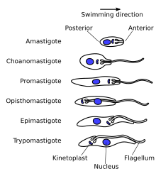
A variety of different morphological forms appear in the life cycles of trypanosomatids, distinguished mainly by the position, length and the cell body attachment of the flagellum. The kinetoplast is found closely associated with the basal body at the base of the flagellum and all species of trypanosomatid have a single nucleus. Most of these morphologies can be found as a life cycle stage in all trypanosomatid genera however certain morphologies are particularly common in a particular genus. The various morphologies were originally named from the genus where the morphology was commonly found, although this terminology is now rarely used because of potential confusion between morphologies and genus. Modern terminology is derived from the Greek; "mastig", meaning whip (referring to the flagellum), and a prefix which indicates the location of the flagellum on the cell. For example, the amastigote (prefix "a-", meaning no flagellum) form is also known as the leishmanial form as all Leishmania have an amastigote life cycle stage.
- Amastigote (leishmanial).[6] Amastigotes are a common morphology during an intracellular lifecycle stage in a mammalian host. All Leishmania have an amastigote stage of the lifecycle. Leishmania amastigoes are particularly small and are among the smallest eukaryotic cells. The flagellum is very short, projecting only slightly beyond the flagellar pocket.
- Promastigote (leptomonad).[6] The promastigote form is a common morphology in the insect host. The flagellum is found anterior of nucleus and flagellum not attached to the cell body. The kinetoplast is located in front of the nucleus, near the anterior end of the body.
- Epimastigote (crithidial).[6] Epimastigotes are a common form in the insect host and Crithidia and Blastocrithidia, both parasites of insects, exhibit this form during their life cycles. The flagellum exits the cell anterior of nucleus and is connected to the cell body for part of its length by an undulating membrane. The kinetoplast is located between the nucleus and the anterior end.
- Trypomastigote (trypanosomal).[6] This stage is characteristic of the genus Trypanosoma in the mammalian host bloodstream as well as infective metacyclic stages in the fly vector. In trypomastigotes the kinetoplast is near the posterior end of the body, and the flagellum lies attached to the cell body for most of its length by an undulating membrane.
- Opisthomastigote (herpetomonad).[6] A rarer morphology where the flagellum posterior of nucleus, passing through a long groove in the cell.
- Endomastigote.[7] A morphotype where the flagellum does not extend beyond the deep flagellar pocket.
Other features[edit]
Notable characteristics of trypanosomatids are the ability to perform trans-splicing of RNA and possession of glycosomes, where much of their glycolysis is confined to. The acidocalcisome, another organelle, was first identified in trypanosomes.[8]
| Trypanosomes | |
|---|---|
 | |
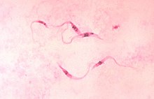
| Trypanosoma cruzi parasites | |
| Scientific classification | |
| Domain: | |
| (unranked): | |
| Phylum: | |
| Class: | |
| Subclass: | |
| Order: | Trypanosomatida Kent 1880 |
| Family: | Trypanosomatidae Doflein 1901 |
| Subfamily | |
| |
https://en.wikipedia.org/wiki/Trypanosomatida#Morphologies


No comments:
Post a Comment