| Trypanosomes | |
|---|---|
 | |
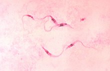
| Trypanosoma cruzi parasites | |
| Scientific classification | |
| Domain: | |
| (unranked): | |
| Phylum: | |
| Class: | |
| Subclass: | |
| Order: | Trypanosomatida Kent 1880 |
| Family: | Trypanosomatidae Doflein 1901 |
| Subfamily | |
| |
Life cycle[edit]
Some trypanosomatids only occupy a single host, while many others are heteroxenous: they live in more than one host species over their life cycle. This heteroxenous life cycle typically includes the intestine of a bloodsucking insect and the blood and/or tissues of a vertebrate. Rarer hosts include other bloodsucking invertebrates, such as leeches,[5] and other organisms such as plants. Different species go through a range of different morphologies at different stages of the life cycle, with most having at least two different morphologies. Typically the promastigote and epimastigote forms are found in insect hosts, trypomastigote forms in the mammalian bloodstream and amastigotes in intracellular environments.
Morphologies[edit]
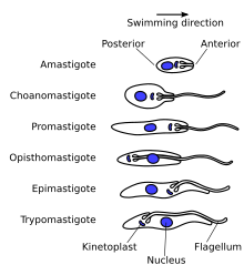
A variety of different morphological forms appear in the life cycles of trypanosomatids, distinguished mainly by the position, length and the cell body attachment of the flagellum. The kinetoplast is found closely associated with the basal body at the base of the flagellum and all species of trypanosomatid have a single nucleus. Most of these morphologies can be found as a life cycle stage in all trypanosomatid genera however certain morphologies are particularly common in a particular genus. The various morphologies were originally named from the genus where the morphology was commonly found, although this terminology is now rarely used because of potential confusion between morphologies and genus. Modern terminology is derived from the Greek; "mastig", meaning whip (referring to the flagellum), and a prefix which indicates the location of the flagellum on the cell. For example, the amastigote (prefix "a-", meaning no flagellum) form is also known as the leishmanial form as all Leishmania have an amastigote life cycle stage.
- Amastigote (leishmanial).[6] Amastigotes are a common morphology during an intracellular lifecycle stage in a mammalian host. All Leishmania have an amastigote stage of the lifecycle. Leishmania amastigoes are particularly small and are among the smallest eukaryotic cells. The flagellum is very short, projecting only slightly beyond the flagellar pocket.
- Promastigote (leptomonad).[6] The promastigote form is a common morphology in the insect host. The flagellum is found anterior of nucleus and flagellum not attached to the cell body. The kinetoplast is located in front of the nucleus, near the anterior end of the body.
- Epimastigote (crithidial).[6] Epimastigotes are a common form in the insect host and Crithidia and Blastocrithidia, both parasites of insects, exhibit this form during their life cycles. The flagellum exits the cell anterior of nucleus and is connected to the cell body for part of its length by an undulating membrane. The kinetoplast is located between the nucleus and the anterior end.
- Trypomastigote (trypanosomal).[6] This stage is characteristic of the genus Trypanosoma in the mammalian host bloodstream as well as infective metacyclic stages in the fly vector. In trypomastigotes the kinetoplast is near the posterior end of the body, and the flagellum lies attached to the cell body for most of its length by an undulating membrane.
- Opisthomastigote (herpetomonad).[6] A rarer morphology where the flagellum posterior of nucleus, passing through a long groove in the cell.
- Endomastigote.[7] A morphotype where the flagellum does not extend beyond the deep flagellar pocket.
Other features[edit]
Notable characteristics of trypanosomatids are the ability to perform trans-splicing of RNA and possession of glycosomes, where much of their glycolysis is confined to. The acidocalcisome, another organelle, was first identified in trypanosomes.[8]
https://en.wikipedia.org/wiki/Trypanosomatida#Morphologies
Excavata is a major supergroup of unicellular organisms belonging to the domain Eukaryota.[1][2][3] It was first suggested by Simpson and Patterson in 1999[4][5] and introduced by Thomas Cavalier-Smith in 2002 as a formal taxon. It contains a variety of free-living and symbiotic forms, and also includes some important parasites of humans, including Giardia and Trichomonas.[6] Excavates were formerly considered to be included in the now obsolete Protista kingdom.[7] They are classified based on their flagellar structures,[5] and they are considered to be the most basal Flagellate lineage.[8]Phylogenomic analyses split the members of the Excavates into three different and not all closely related groups: Discobids, Metamonads and Malawimonads.[9][10][11] Except for Euglenozoa, they are all non-photosynthetic.
https://en.wikipedia.org/wiki/Excavata
Kinetoplastida (or Kinetoplastea, as a class) is a group of flagellated protists belonging to the phylum Euglenozoa,[3][4] and characterised by the presence of an organelle with a large massed DNA called kinetoplast (hence the name). The organisms are commonly referred to as "kinetoplastids" or "kinetoplasts"[5] The group includes a number of parasites responsible for serious diseases in humans and other animals, as well as various forms found in soil and aquatic environments. Their distinguishing feature, the presence of a kinetoplast, is an unusual DNA-containing granule located within the single mitochondrion associated with the base of the cell's flagellum (the basal body). The kinetoplast contains many copies of the mitochondrial genome.
The kinetoplastids were first defined by Bronislaw M. Honigberg in 1963 as the members of the flagellated protozoans.[1]They are traditionally divided into the biflagellate Bodonidae and uniflagellate Trypanosomatidae; the former appears to be paraphyletic to the latter. One family of kinetoplastids, the trypanosomatids, is notable as it includes several genera which are exclusively parasitic. Bodo is a typical genus within kinetoplastida and including various common free-living species which feed on bacteria. Others include Cryptobia and the parasitic Leishmania.
| Kinetoplastida | |
|---|---|
 | |
| Trypanosoma cruzi parasites |
https://en.wikipedia.org/wiki/Kinetoplastida
A kinetoplast is a network of circular DNA (called kDNA) inside a large mitochondrion that contains many copies of the mitochondrial genome.[1][2] The most common kinetoplast structure is a disk, but they have been observed in other arrangements. Kinetoplasts are only found in Excavata of the class Kinetoplastida. The variation in the structures of kinetoplasts may reflect phylogenic relationships between kinetoplastids.[3] A kinetoplast is usually adjacent to the organism's flagellar basal body, suggesting that it is tightly bound to the cytoskeleton. In Trypanosoma brucei this cytoskeletal connection is called the tripartite attachment complex and includes the protein p166.[4]
https://en.wikipedia.org/wiki/Kinetoplast


No comments:
Post a Comment